Medical Device Company

Paracentesis Needle

Next Image Title

Artist Name

A patient with ascites, during which a paracentesis procedure is performed to drain fluid from the peritoneal cavity.

Mike Austin

AMI #28

Page 70

AMI #27

Page 64

AMI #26

Page 60

AMI #25

Page 68

AMI #24

Page 67

AMI #23

Page 83

AMI #22

Page 18

AMI #21

Page 19

AMI #20

Page 20

medical illustration of This illustration highlights the occupational risks faced by healthcare professionals working in fluoroscopic laboratories. Prolonged exposure to ionizing radiation places clinicians at increased risk for cancer, cataracts, and reproductive health concerns. In addition, the long-term use of heavy lead aprons—while essential for radiation protection—contributes to chronic orthopedic strain and musculoskeletal injury.
medical illustration of This medical illustration presents the vascular anatomy of the upper extremity with detailed insets highlighting common arteriovenous (AV) fistula configurations and surgically transposed vessels used to establish hemodialysis access. The primary image depicts the arterial and venous structures of the arm in anatomical context, while enlarged inset panels demonstrate specific fistula locations and vessel transpositions with precision and clarity.
medical illustration of This five-step illustration of lupus cell therapy visually explains the process of immune cell collection, modification, and reinfusion to treat autoimmune disease. Designed for clarity and accuracy, it transforms complex immunology into an accessible and engaging visual.
medical illustration of This illustration highlights a catheter insertion site in the chest, emphasizing both anatomical accuracy and infection prevention strategy. The catheter is shown entering the skin and advancing into the underlying vasculature, with careful attention to surrounding tissue layers and placement. At the point of entry, a specialized antimicrobial gel is applied over the skin surface, demonstrating its role as a protective barrier against bacterial contamination.
medical illustration of This medical illustration depicts a drainage catheter positioned within the peritoneal cavity for the treatment of ascites. Key abdominal anatomy is shown in context, with fluid accumulation clearly visualized within the abdominal cavity to demonstrate the pathological condition. The illustration highlights catheter placement relative to surrounding structures, helping convey both the clinical purpose of the device and the anatomical environment in which it functions.
medical illustration of A TIPS (Transjugular Intrahepatic Portosystemic Shunt) procedure is a minimally invasive treatment for portal hypertension, a condition where blood pressure in the portal vein is abnormally high, often due to liver disease.
medical illustration of Depiction of a stent graft treating atherosclerotic disease in a peripheral artery.
medical illustration of Peroneal tendon repair using sutures and a bone marrow aspirate implant.
medical illustration of After the creation of an A-V fistula, blood flow circulates down the veins to revascularize the toes and foot and creating collateral vessels.
medical illustration of Infographic of a tree and root system is a representation of psoriatic disease as an immune-mediated inflammatory disease with underlying systemic inflammation contributing to various comorbidities, including hidden in the roots, cardiovascular disease, obesity, mental health conditions and psoriatic arthritis.
medical illustration of Various research journal cover illustrations supporting articles.
medical illustration of Illustration depicting the relationship of salivary glands, facial arteries and veins.
medical illustration of Illustration depicting the relationship of salivary glands and muscles.
medical illustration of Liver tumor injection of a substance to cause an antibody response to activate T-cells and attack cancer cells.
medical illustration of This medical illustration depicts a patient with ascites undergoing a paracentesis procedure to drain excess fluid from the peritoneal cavity. The image highlights relevant abdominal anatomy and fluid accumulation, while focusing on the specialized drainage catheter. The catheter features a unique safety tip that allows the needle to extend upon contact for controlled access, then automatically retract without tension, reducing the risk of tissue injury.
medical illustration of Part of a surgical guide to depict reverse total shoulder arthroplasty.
medical illustration of Pulmonary arteriovenous malformations in the lung with occlusion devices. Artery, vein, vascular
medical illustration of Achilles tendon repair technique guide.
medical illustration of This medical illustration depicts a breast cancer patient undergoing radiation therapy traveling through a metal free port. The patient is shown lying in a treatment position while a focused external radiation beam passes safely through the port, allowing uninterrupted delivery of radiation to a tumor located beneath it.
medical illustration of This illustration of a dental implant was created for product marketing.  Replacement tooth roots, oral health, prosthetic replacement, osseointegration, jawbone, titanium, endosteal, subperiosteal
medical illustration of Part of a series of illustrations depicting hypoplastic left heart syndrome (HLHS) and repair created for a Children's Hospital. Birth defect, blood flow, congenital, baby, surgery, left ventricle, mitral valves, aortic valve, aorta, atrial septal defect
medical illustration of Stent placed in the iliac-femoral vein with blood flow. Disease, thrombus, peripheral disease, pelvis,
medical illustration of Illustration depicting surgically targeted radiation tiles placed in brain tissue for treating brain tumors.
medical illustration of Flexor digitorum longus tendon transfer in the foot. Toes, ankle, posterior tibial tendon (PTT), navicular bone, surgical treatment, screw fixation
medical illustration of A device that depicts the removal of instent restenosis and thrombus. Peripheral artery disease, PAD, stent, vascular, plaque, atherosclerosis
medical illustration of Drug eluting balloon in the femoral artery of the leg.  Disease, peripheral, PAD, knee, hip, pelvis,
medical illustration of Illustration of a femoral bone lesion biopsy.
medical illustration of Treatment for benign prostatic hyperplasia supporting a journal article.
Enlarged, prostate, gland
medical illustration of Thumb carpal-metacarpal arthritis repair using the ATLAS device after removal of the trapizium bone.  Hand, joint, wrist,
medical illustration of A very small angioplasty balloon for patients with disease that is in vessels below the knee.  Peripheral artery disease, PAD, leg, atherosclerosis
medical illustration of Central venous catheter placement into the subclavian vein and into the superior vena cava.  Heart, chest, port,
medical illustration of This medical illustration depicts a self-expanding,  vascular embolization device deployed within the ovarian vein for the treatment of pelvic congestion syndrome.
The illustration is designed to support patient education, clinical communication, and medical device marketing by clearly visualizing device function and anatomical context.
medical illustration of Occlusion device placed in the gastroduodenal artery. The illustration includes the stomach, pancreas, liver, and gall bladder.
medical illustration of Artery-vein loop graft in the arm.
medical illustration of Blood flow and capture of thrombus in a vena cava filter.  Kidneys, renal, IVC, aorta, vein, artery.
medical illustration of Pelvic section illustrating female anatomy, including the uterus, bladder, vagina, colon and clitorus, with a vaginal wall close-up.
medical illustration of This visual illustrates venous disease in the iliac vein with post balloon dilation of the stent.  Pelvis, pelvic, femoral, PAD,
medical illustration of Drug eluting balloon treating peripheral artery disease.  Angioplasty, PAD, balloon catheter,
medical illustration of This illustration was created for advertising/marketing and depicts blood flow through a flared arteriovenous graft.
medical illustration of Final illustration of an ankle syndesmosis series depicting the repair of a high ankle sprain and fibular fracture.
medical illustration of Cover illustration supporting an article on Hypertrophic Cardiomyopathy of the heart in youth sports.
medical illustration of Patient education illustration showing hemodialysis, peritoneal dialysis and arteriovenous graft.
medical illustration of A thyroid stimulating hormone is released from the pituitary gland which causes the release of another hormone.
medical illustration of Illustration in support of a journal article depicting Interstitial cystitis or painful bladder syndrome.
medical illustration of Valvuloplasty perfusion catheter that allows blood flow while the balloon is inflated.  Heart, aortic valve, aorta,
medical illustration of Device that measures blood volume to assess the probability of peripheral artery disease.
medical illustration of Radiofrequency ablation catheter collapsing the saphenous vein. Pelvis, valve, ablation, RF, reflux, heat, peripheral, disease
medical illustration of Marketing illustration for an implantable port located under the skin, over the ribs and in the peritoneum.
medical illustration of Balloon angioplasty for peripheral artery disease. PAD, catheter, leg, introducer, atherosclerosis

Profile

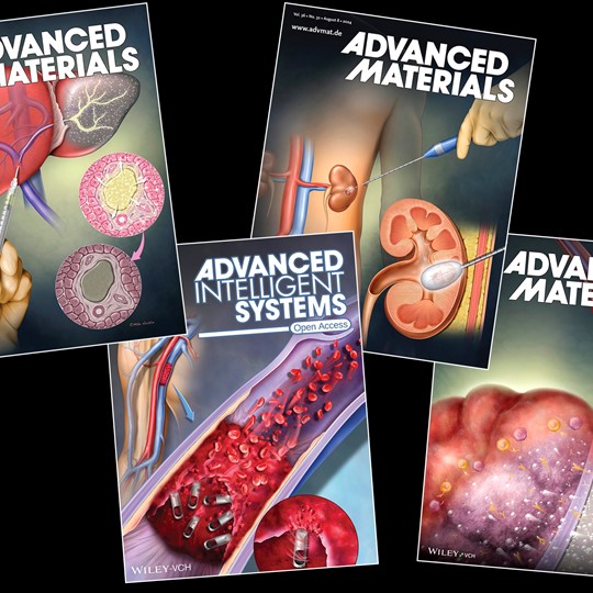
Mike Austin, owner of Internal Art Medical Illustration, is known for his skillful creativity and artistic style. Mike is an independent medical illustrator with over two decades of experience collaborating with clients and creating compelling visuals to communicate information clearly. His intent is to create a warm, appealing and accurate final illustration that is delivered on time. In 1994, Mike graduated with a Master’s degree in Medical and Biological Illustration from the University of Michigan School of Medical Illustration in Ann Arbor, Michigan. His background includes rigorous coursework in gross anatomy, comparative anatomy, pathology and biology. With his expertise in science, anatomy and artistry, Mike can find a solution to your intricate communication goals. Prior to starting Internal Art Medical Illustration, Mike began his career at the Arizona Heart Institute in Phoenix, Arizona. He is a professional member of the Association of Medical Illustrators since 1999. Many clients have mentioned they appreciate the flexibility and timeliness that Mike provides when working on their projects. His work has appeared in PBS documentaries, medical malpractice trials, surgical and clinical journals, books, advertising, professional and patient education materials and used for medical device marketing and packaging.

Style/Techniques

Airbrush, Color, Information Graphics

Subject/Specialties

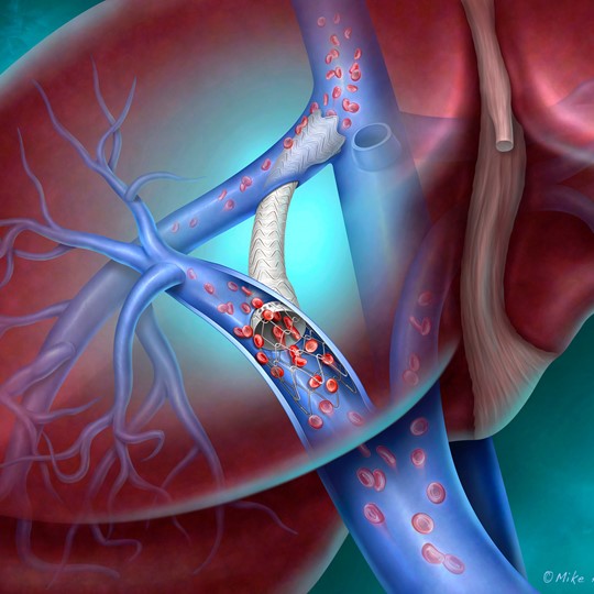
Allergy / Immunology, Alternative Medicine, Anatomy, Biotechnology, Cardiac Surgery / Cardiology, Cell biology / Histology, Dentistry, Dermatology, Disease Management, Endocrinology / Metabolic, Gastroenterology, General Surgery, Maternal / Child, Medical Devices, Molecular Biology, Neuroscience, Neurosurgery, Obstetrics / Gynecology, Orthopaedics, Pediatrics, Physical Therapy, Reconstructive Surgery, Reproductive Biology, Respiratory, Thoracic Surgery, Urology, Vascular Surgery, Virology, Injuries, Oncology, Mechanism of Action (MOA), Health & Wellness, Sports Medicine