SerbinCreative

(805) 963-0439 | Toll-Free (800) 876-6425

www.serbincreative.com LA / NY / UK

Mark M. Miller

medical illustration of Editorial. View of young girl suffering an epileptic seizure. Brain with electrical storm, EEG and seizure medication
medical illustration of Editorial. Woman being palpated for thyroid tumor. Viw of iodine scan, blood workup, thyroid and antibodies
medical illustration of Editorial. View of man suffering from flank pain. View of kidney with stone, stone undergoing laser ablation.
medical illustration of Editorial. View of man coughing with diseased lungs in background. View of bacteria and isoniazid
medical illustration of Editorial. Montage illustration of carpal tunnel injury and treatment
medical illustration of Editorial. Montage illustration demonstrating feet ulcers due to diabetes. Includes compromised leg circulation, ulcer care and revascularization procedure
medical illustration of Editorial. Montage showing a woman in distress with imagery of a breast with tumor, and a breast exam
medical illustration of Editorial.
medical illustration of Editorial. Montage image showing older woman rubbing her disfigured hands and neck. Methotrexate shown, plus antiboides, lymphocytes
medical illustration of Editorial. Montage illustration showing a baseball pitcher throwing a ball, with other images showing a rotator cuff tear and injection
medical illustration of Editorial. Montage illustration depicting a man smoking cigarette, man coughing and undergoing breathing study due to COPD. A diseased lung alveoli is included.
medical illustration of Editorial. Montage illustration showing man with bad sunburn, and same man being examined by a dermatologist. Includes inset of skin lesion, skin biopsy and fluorouracil application.
medical illustration of Editorial. Montage illustraton depicting imagery related to lower gastrointestinal disease, specifically crohn's disease. Show a woman with flank pain, the ileocecal junction with disease, and genetic elements
medical illustration of Editorial. Montage illustratration depicting elements related to infectious disease. Shows man with headache, viruses, blood borne disease and vectors such as mosquito and a bird
medical illustration of Editorial. Montage illustration depicting visuals related to diabetes. Shows an overweight man, glucose measurement device, diabetic retinopathy, pancreas and medication
medical illustration of Editorial. Montage illustration showing imagery related to valvular heart disease. Includes a man with chest pain, and EKG, a diseased valve and balloon angioplasty plus stent
medical illustration of Editorial. Montage illustration depicting imagery related to heart disease, including smoking, sodium, blood pressure, diet. Includes image of coronary artery plaque and statin
medical illustration of Editorial. Illustration showing two feet on floor scale. The scale indicates diseases related to obesity, rather than weight.
medical illustration of Editorial. Montage illustration showing imagery related to pregnancy care, including a sonogram and blood testing
medical illustration of Editorial. Illustration showing obese woman and health professional taking her blood pressure. Includes imagery of artery, RBCs and cellular processes
medical illustration of Four medical illustrations demonstrating the anatomy of the kidneys: Placemant in the body, an external view, a cross-sectional view, and an illustraiton of the nephron
medical illustration of Mid-sagittal and anterior views of the human skull, with individual bones color coded
medical illustration of Highly detailed medical illustration showing the human brain in frontal section with the motor cortex highlighted as well as it’s blood supply
medical illustration of A medical illustration showing a full term baby in breech position
medical illustration of A medical illustration detailing the anatomy of the diaphragm and stomach from an anterior view. With labels.
medical illustration of A medical illustration showing birth stations from an anterior perspective
medical illustration of An anteror view of the knee with labels
medical illustration of A lateral view of the knew with a scope inserted into the knee joint
medical illustration of Anatomic view of the middle ear. Coronal section through ear, ear canal and eardrum showing ossicles and cochlea
medical illustration of Three cross-section medical illustrations demonstrating injuries to 3 separate lumbar joint spaces, with protruding disks pressing on spinal nerve roots. With spinal stenosis and joint hypertrophy
medical illustration of Realistic rendering of 7 human cataracts
medical illustration of A medical illustration showing  a human eye and an intraocular lens (IOL) in the process of being place in the eye
medical illustration of A view of a kidney with late stage polycystic kidney disease and a diseased nephron
medical illustration of Two surgical illustrations for a textbook detailing steps in the The Middle Cranial Fossa Approach to Vestibular Schwannomas
medical illustration of Three surgical illustrations for a texbook detailing steps in the subtemporal approach to the skull base
medical illustration of A series of medical illustrations for publication showing in the fusion of the lumbar fourth and fifth lumbar verterbrae using bone chips, a fixation device and bone cages
medical illustration of A medical illustration for a patient education brochure detailing a Chait Percutaneous Cecostomy Catheter in situ in the cecum of the colon
medical illustration of A medical illustration of an eye in cross-section showing a ray of light entering the pupil and being sharply focused on the fovea centralis of the retina
medical illustration of An inside scientific illustration for a corporate report showing the development of the sea anemone nematostella vectensis with an emphasis on HOX gene expression and radial symmetry
medical illustration of Inside page scientific illustration for corporate report detailing the complex migration of neural crest cells and the resulting tissues in the body that derive from these cells.
medical illustration of An illustration of the lifecycle of a flea shown in 4 stages
medical illustration of Side views of the blind cave fish Astyanax mexicanus, and it's sighted surface-dwelling relative
medical illustration of A veterinary illustration for publication in two parts which detail a dogs partotid glands and cranial nerves.
medical illustration of A wall chart showing the anatomy and lifecycle of the fruit fly drosophila melanogaster. For a lay audience.
medical illustration of A scientific and marketing illustration for the veterinary pharmaceutical product Lymevax®
medical illustration of A veterinary illustration for presentation depicting surgery on a cat to place a esophagostomy tube while avoiding important structures
medical illustration of A veterinary illustration for a trade show product showing canine tapeworm inside the intestine of a dog. To be used to advertise deworming pharmaceutical
medical illustration of Fine art rendering of the Atlas Moth
medical illustration of Fine art rendering of a praying mantis camouflaged in shrubbery.
medical illustration of Fine Art rendering of the butterfly Zalmoxis

Profile

Mark Mitchell Miller, owner of Miller Medical Illustration and Mark Miller Creations, was born and raised in Kansas City, Missouri. An early interest in both art and science led him to combine a Bachelor of Fine Arts degree and Pre-Medicine at the University of Missouri, Columbia. Working with Dr. Robert Breitenbach in the Department of Biology his senior year, Mark illustrated an undergraduate lab manual, a project which switched his focus from a career in medicine to one in medical illustration. Mark subsequently entered the graduate program in Medical Illustration at the Department of Art as Applied to Medicine, The Johns Hopkins University School of Medicine, Baltimore. His interests there included 3D medical models as well as facial prosthetics. Upon receiving a Master of Arts degree in Medical Illustration in 1986, Mark joined the faculty in Art as Applied to Medicine, where his responsibilities included teaching medical sculpture, heading the facial prosthetics clinic, and producing surgical illustrations for the Department of Otolaryngology, Head and Neck Surgery. In 1991 Mark established Miller Medical Illustration, a sole-proprietorship, in Kansas City. In July of 2013 he joined the Stowers Institute for Medical Research, Department of Communications, where he is employed on a part-time basis as Scientific Illustrator and Instructor in Adobe Illustrator and Best Practices in Visual Communications. Mark continues to create medical and scientific illustrations for a diverse group of national and international clients, depicting a wide range of medical, biological, scientific and veterinary subjects, and is a speaker on the field of medical illustration and its history.

Style/Techniques

Airbrush, Black & White, Color, 3D

Subject/Specialties

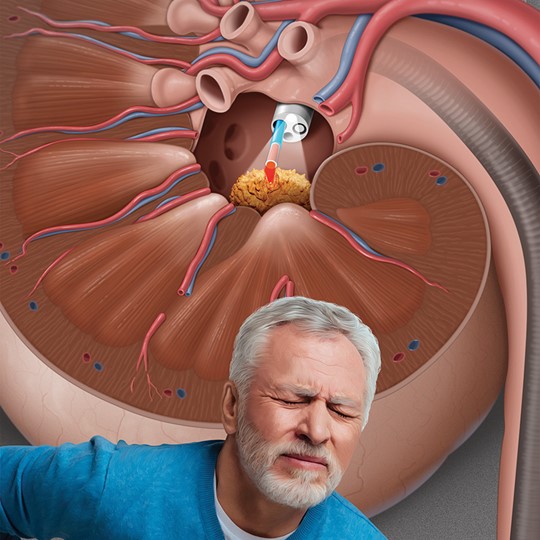
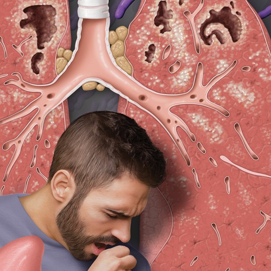
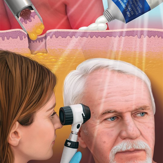
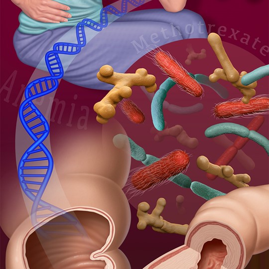
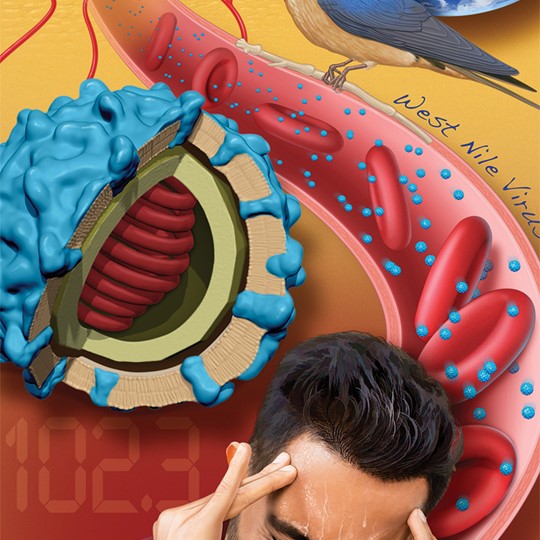
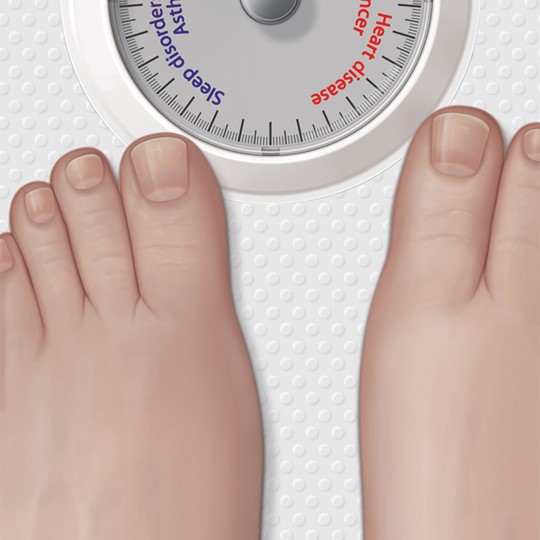
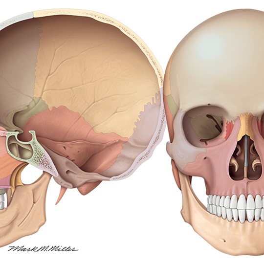
Allergy / Immunology, Anatomy, Biology, Biotechnology, Botany, Cardiac Surgery / Cardiology, Cell biology / Histology, Dentistry, Dermatology, Disease Management, Embryology, Emergency Medicine, Endocrinology / Metabolic, Entomology, Gastroenterology, General Medicine, General Surgery, Genetics, Maternal / Child, Medical Devices, Natural History, Natural Science / Nature, Neuroscience, Neurosurgery, Obstetrics / Gynecology, Ophthalmology, Orthopaedics, Pediatrics, Pharmacology, Reconstructive Surgery, Reproductive Biology, Respiratory, Thoracic Surgery, Transplantation Surgery, Urology, Vascular Surgery, Veterinary Medicine, Virology, Zoology, Injuries, Legal Exhibits, Oncology, Health & Wellness, Pathology, Plastic Surgery, Sports Medicine