Histology of the mouse prostatic duct
Share:


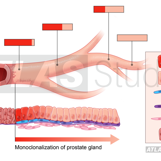


This illustration shows the different cell types in the mouse prostatic duct. Each cell can be edited separately, so different cells can be highlighted for different focuses of the client's research of the mouse prostate. mouse prostate, animal research, cancer research, pharmacology, physiology, toxicology, histology, duct, anatomy, cell types, cell division, epithelium, basal layer, visual abstract, urology
Keywords: Color, Line with Color, Research, Anatomy, Biology, Cell biology / Histology, Urology
© Roswell Park